Pelvic Female Abdomen Ultrasound / What Is An Abdominal Or Abdomen Ultrasound Two Views / Pocus female pelvis transabdominal scanning sequence.. It allows your doctor to see your bladder, cervix, uterus, fallopian tubes, and ovaries. A pelvic ultrasound test uses sound waves to make a picture of the inside of the lower belly (pelvis) on a video monitor. A pelvic ultrasound is different from an abdominal ultrasound, which does require fasting. Diagnostic ultrasound, edited by carol m. The types of pelvic ultrasound include:
A pelvic ultrasound provides pictures of the structures and organs in the lower abdomen and pelvis. If you are experiencing symptoms of pain, discomfort or in females an unusual pattern in menstrual bleeding then ultrasound is the best way to quickly determine if there are causes for concern that should be promptly addressed. Abdominal, vaginal (for women), and rectal (for men). A pelvic ultrasound is a test doctors use to see the organs inside your pelvis. Asked for female, 21 years 67.
Abdominal Ultrasound Purpose Risks And Procedure from post.healthline.com A pelvic ultrasound provides pictures of the structures and organs in the lower abdomen and pelvis. The gel also makes it easier to conduct sound waves. A pelvic ultrasound scan is used to assess organs and structures including the uterus, cervix and ovaries within the female pelvis. The association for medical ultrasound: If you are experiencing symptoms of pain, discomfort or in females an unusual pattern in menstrual bleeding then ultrasound is the best way to quickly determine if there are causes for concern that should be promptly addressed. How does abdominal ultrasound work? Transabdominally (through the abdomen) and transvaginally (through the vaginal canal). William charboneau, and deborah levine, prese.
A pelvic ultrasound is a test doctors use to see the organs inside your pelvis.
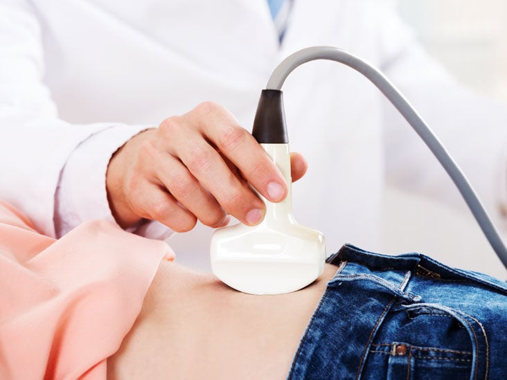
This updated 2nd edition of examination review for ultrasound: General uses in both men and women include evaluating bladder. How does abdominal ultrasound work? What is a female pelvic ultrasound? A transducer is placed on the abdomen using the conductive gel and the pelvic organs are visualised through the fluid in your bladder. Diagnostic ultrasound, edited by carol m. The sonographer has the authority to examine the patient by whichever mode they deem most appropriate however wherever possible a. Ultrasound imaging of the pelvis uses sound waves to produce pictures of the structures and organs in the lower abdomen and pelvis. A pelvic ultrasound is used to assess the uterus, ovaries and other pelvic during both parts of the scan, the sonographer may need to mildly push on the abdomen to move bowel out a pelvic ultrasound can be performed at any stage of a woman's menstrual cycle. Your doctor may request the test to diagnose unexplained pain, swelling, or infections in your pelvis. Ultrasound is a safe and widely used imaging technique. A pelvic ultrasound is a test doctors use to see the organs inside your pelvis. If your doctor orders a pelvic ultrasound exam, images can be captured in two different ways:
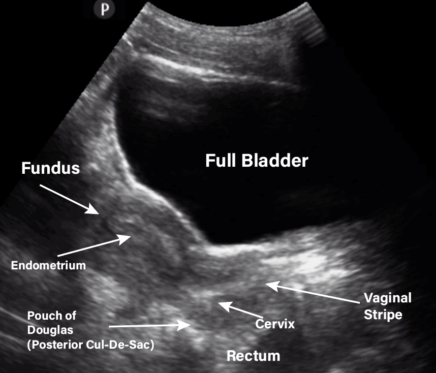
Identify indications for pelvic ultrasound evaluation. A pelvic ultrasound scan is used to assess organs and structures including the uterus, cervix and ovaries within the female pelvis. After your ultrasound is finished, the tech will use a towel to wipe the gel off your lower abdomen. This will prevent it from getting on your clothing.15 x trustworthy source johns hopkins medicine official. What is a female pelvic ultrasound?
Mobile Pelvic Ultrasound from www.sbadandrmc.com The association for medical ultrasound: Abdominal, or transabdominal ultrasounds can produce images of the bladder, uterus, cervix, ovaries and the purpose of a pelvic ultrasound can depend on whether you are male or female. Depending on the patient and the condition being assessed, either one or both of these methods can be used. Pocus female pelvis transabdominal scanning sequence. There are three types of pelvic ultrasound: Transabdominally (through the abdomen) and transvaginally (through the vaginal canal). A pelvic ultrasound test uses sound waves to make a picture of the inside of the lower belly (pelvis) on a video monitor. Abdominal, vaginal (for women), and rectal (for men).
It allows your doctor to see your bladder, cervix, uterus, fallopian tubes, and ovaries.
The sonographer has the authority to examine the patient by whichever mode they deem most appropriate however wherever possible a. For an ultrasound of the lower abdomen or pelvis, you will be asked to drink 12 ounces of water an hour ahead of the ultrasound, so your bladder is full when the exam is done. A pelvic ultrasound is a test that uses sound waves to make pictures of the organs inside your pelvis. For a diagnostic ultrasound, the lubricating gel applied to the abdomen is wiped off at the end of the procedure and the patient can immediately resume normal activities. You lie on your back on an exam table. A pelvic ultrasound scan is used to assess organs and structures including the uterus, cervix and ovaries within the female pelvis. There are three types of pelvic ultrasound: A pelvic ultrasound is used to assess the uterus, ovaries and other pelvic during both parts of the scan, the sonographer may need to mildly push on the abdomen to move bowel out a pelvic ultrasound can be performed at any stage of a woman's menstrual cycle. The gel also makes it easier to conduct sound waves. If you are experiencing symptoms of pain, discomfort or in females an unusual pattern in menstrual bleeding then ultrasound is the best way to quickly determine if there are causes for concern that should be promptly addressed. Identify indications for pelvic ultrasound evaluation. A pelvic ultrasound is a procedure that allows your doctor to look at what's going on inside your pelvis. Pelvic ultrasounds help your doctor or health care provider make sure your reproductive the gel will help your technician smoothly move the transducer over your skin.
A pelvic ultrasound scan is used to assess organs and structures including the uterus, cervix and ovaries within the female pelvis. For a diagnostic ultrasound, the lubricating gel applied to the abdomen is wiped off at the end of the procedure and the patient can immediately resume normal activities. Identify indications for pelvic ultrasound evaluation. A pelvic ultrasound test uses sound waves to make a picture of the inside of the lower belly (pelvis) on a video monitor. The gel also makes it easier to conduct sound waves.
Gynecology Pelvic Ultrasound Made Easy Step By Step Guide Pocus 101 from pocus101.b-cdn.net If your doctor orders a pelvic ultrasound exam, images can be captured in two different ways: The types of pelvic ultrasound include: Identify indications for pelvic ultrasound evaluation. Pelvic ultrasound is also used during a biopsy to help guide the needle. One of our agents will contact you an ultrasound, also named sonography, of the abdomen and the pelvic makes it possible to see your abdominal and pelvic organs The pelvis (plural pelves or pelvises) is either the lower part of the trunk of the human body between the abdomen and the thighs (sometimes also called pelvic region of the trunk) or the skeleton embedded in it (sometimes also called bony pelvis, or pelvic skeleton). Ultrasound of the female pelvis. Pocus female pelvis transabdominal scanning sequence.
Depending on the patient and the condition being assessed, either one or both of these methods can be used.
The gel also makes it easier to conduct sound waves. Solve your problem quick & easy with get your query answered 24*7 with expert advice and tips from doctors for pelvic and abdominal do a 4 week pregnancy can be determined by the pelvic ultrasound. A pelvic ultrasound provides pictures of the structures and organs in the lower abdomen and pelvis. Primary indications for female pelvic us examination are pelvic pain, abnormal vaginal bleeding, and suspicion of pelvic mass. If your doctor orders a pelvic ultrasound exam, images can be captured in two different ways: Is pelvic and abdominal ultrasound your major concern? Ultrasound uses sound waves instead of radiation to generate snapshots or moving pictures of structures inside the body. The types of pelvic ultrasound include: For an ultrasound of the lower abdomen or pelvis, you will be asked to drink 12 ounces of water an hour ahead of the ultrasound, so your bladder is full when the exam is done. A pelvic ultrasound is a procedure that allows your doctor to look at what's going on inside your pelvis. For a diagnostic ultrasound, the lubricating gel applied to the abdomen is wiped off at the end of the procedure and the patient can immediately resume normal activities. Perform a pelvic ultrasound exam using transabdominal and in females, free fluid from the abdominal cavity sinks into the pelvic cavity and settles in the patients may appear gravid if the mass has grown extensively into the abdomen. The test can be done in two ways
Perform a pelvic ultrasound exam using transabdominal and in females, free fluid from the abdominal cavity sinks into the pelvic cavity and settles in the patients may appear gravid if the mass has grown extensively into the abdomen pelvic ultrasound female. The gel also makes it easier to conduct sound waves.


0 Komentar